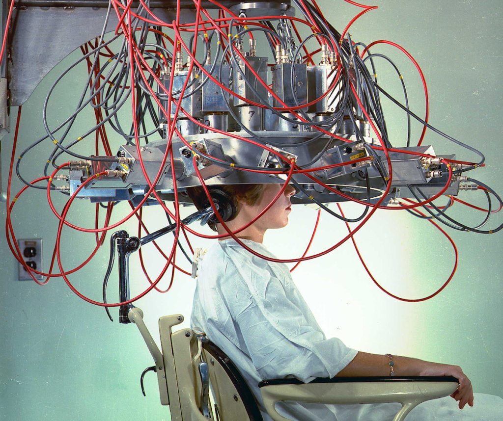
You’ve probably seen a TV show or read articles that feature impressive brain imagery depicting a “normal” brains vs. depressed, anxious, or addicted brains.
Brain scans have made huge improvements in diagnosis and treatment.
However critics complain that when these images are interpreted to reveal information about your thoughts, feelings, and cognition, the claims are often exaggerated.
A recently published study by M.I.T.’s Edward Vul (and associates) concluded that,
“a disturbingly large, and quite prominent, segment of fMRI research on emotion, personality, and social cognition is using seriously defective research methods and producing a profusion of numbers that should not be believed.”
Brain scans were developed in order to visualize brain structures. Now they are being used to interpret brain function. This is where we need to proceed with caution. Personally I’ve had misgivings about who is deciding what constitutes a “normal” brain.
Would Beethoven’s or Einstein’s brain have lived up to the test?
And what about the invasive nature of such technology? Body scanners at airport security have been in the headlines. If you don’t like the idea of your body being displayed for all to see, how would you feel about your brain being scanned and interpreted without your permission?
NPR reports the US military is developing portable brain scanners that work from a distance and can supposedly determine whether a person is lying or telling the truth. While this might be useful at security checkpoints, the ramifications of such technology are disturbing, especially if the software (or human being) that is interpreting such a brain scan is less than 100% accurate.
Clearly we need some ethical guidelines is place as this technology emerges.
Brain scans aren’t going away. Nor should they. They are powerful, life-saving tools. But perhaps we need to proceed with caution about how they are being interpreted, and “sold” to the public outside of medical purposes. Especially when brain scans are being used to interpret intentions, emotions, and feelings.
In an article for Scientific American Mind, Michael Shermer (publisher of Skeptic Magazine) described the limits of fMRI (functional magnetic resonance imaging). Here’s a summary:
1. Studies Are Skewed by Selection and Environment
The population sample can’t be truly random because about 20% of experiment subjects back out because of the claustrophobic experience of being in a narrow tube with the head locked firmly in place while being subjected to the loud banging of the machine. All this, while attempting to watch images, make choices, and report on emotions. It’s obvious that the brains monitored under such conditions are unlikely to reflect “real world” conditions.
2. Scans Don’t Measure Direct Brain Activity
It’s an oversimplification to describe parts of the brain “lighting up” when a person is thinking about something specific. It’s important to remember that an fMRI isn’t showing actual mental activity. It’s indirectly interpreting such activity by measuring the flow of blood to an area of the brain. Blood flow is known to take longer to register than the time it takes to think the thoughts that supposedly create the increased blood flow.
3. The Pretty Colors Are Fake
Such computer-generated images give the false impression that a certain well-defined area of the brain is responsible for specific activities. It’s much more likely that neural activity is distributed in a more loosely defined network. Another point to keep in mind according to Shermer: Scientists know that most brain activity is not stimulus driven. It’s occurring spontaneously all the time. Therefore the task to separate out which part of the brain is performing a specific task at any given moment is a challenge.
4. You’re Not Looking at a Portrait of One Brain
The images you’ve seen are probably not one person’s brain. Most likely the image was created from a statistical compilation of all the brain scans in a study. Even the brain scan image of one brain is actually a result of computer-adjusted data, using software-generated corrections for head movement as well as other possible variables. It’s not as simple as a camera taking a picture.
5. Interpreting Brain Scans Is an Art, Not a Science
Here’s an example: Typically, a structure in the brain called the amygdala is associated with a fear response. But the amygdala is also activated in response to arousal and even positive emotions. So if your amygdala “lights up” in a brain scan, it doesn’t necessarily mean you’re experiencing fear. Similarly, the right pre-frontal cortex lights up when focusing on certain specific tasks, but it’s also involved when you do almost any difficult task.
A Glossary of Brain Scans:
X-ray: Discovered in 1895 by Wilhelm Conrad Roentgen who received the first Nobel Prize for his work. Electromagnetic radiation passes through an object projecting an image onto light-sensitive film. Modern digital x-ray uses less radiation.
EEG (electroencephalograph. Multiple electrodes attached to the scalp provide a direct reading of the electrical activity in your brain. In use since the 1920’s. It’s non-invasive but produces a chart and not an image. It also can’t record activity in the deeper areas of the brain.
CAT Scan (computed axial tomography) Also called a CT scan (computed tomography) Used since the 1970’s, uses special x-ray equipment and computers in order to visualize areas in sections that would not be visible in a normal x-ray.
PET (positron emission tomagraphy) Measures blood flow in different parts of the brain by tracking a small amount of radioactive substance which is given to the patient and detected by special cameras.
MRI (magnetic resonance imaging) Magnetic fields are used to generate computer images of internal structures.
fMRI (functional magnetic resonance imaging) Similar to MRI but able to measure and monitor brain activity and blood flow in real time.
MEG (magnetoencephalograpy) Detects real time brain activity by measuring magnetic fields that are created by electric current flowing within neurons.
SPECT (single photon emission computed tomography) Similar to a PET, uses a radioactive tracer to monitor blood flow in the brain. Produces a three dimensional image.
DTI (diffusion tensor imaging) Measures the flow of water molecules along myelin which makes up 50% of the brain. This technology is not yet easily interpreted.
The source material for this article is from the excellent and thoughtful book The Scientific American Brave New Brain, How Neuroscience, Brain-Machine Interfaces, Neuroimaging, Psychopharmacology, Epigentics, the Internet, and Our Own Minds Are Stimulating and Enhancing the Future of Mental Power
Related Articles:
Your Brain Is Plastic – Is That a Good Thing?
Some Random Notes On Memory

